

Files
Download Full Text (599 KB)
Description
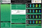
CT Perfusion (CTP) scanning of the brain is an essential part of the currently used diagnostic algorithm in the management of patients presenting with acute stroke. The current stroke imaging workup is initiated with a noncontrast head CT to rule out a possible hemorrhagic cause of the stroke symptoms or an intracranial hematoma which would represent a contraindication for intravenous lysis treatment. According to the current literature, CTP scans of the brain are of particular benefit for stroke patients arriving within 5-24 hours following the onset of symptoms or with unknown onset of symptoms such as wake-up strokes, which are unlikely to benefit from intravenous thrombolytic treatment, but will potentially benefit from intra-arterial thrombolysis or thrombectomy. CTP is used, ultimately to determine the total amount of ischemic cerebral tissue and to estimate the amount of salvageable cerebral tissue, to determine a potential benefit from endovascular therapy. The interpretation of CTP images is based on the evaluation of different perfusion parameter maps. The cerebral perfusion maps [1] include cerebral blood volume (CBV), cerebral blood flow (CBF), and perfusion delay maps, with mean transit time (MTT) and time to peak (TTP) maps most commonly used. A perfusion defect on CBV with perfusion defects matching in size and location on CBF and MTT or TTP maps indicate nonsalvageable cerebral tissue. In case of a normal CBV map, perfusion defects on MTT/TTP and CBF maps represent cerebral tissue at risk for infarct or salvageable cerebral tissue. Perfusion defects that are matching on all perfusion maps to include CBV are also referred to as core infarct i.e. non-salvageable tissue, as opposed to possible areas of salvageable cerebral tissue adjacent to the core infarct, called penumbra. Patients with a significant amount of penumbra with smaller core are candidates for endovascular therapy.
Publication Date
Summer 2020
Publisher
Xavier University of Louisiana
City
New Orleans
Keywords
CT Perfusion, Imaging, Brain
Disciplines
Medical Education
Recommended Citation
Nguyen, Henry and Lammle, Markus, "Introduction to CT Perfusion Imaging of the Brain." (2020). Building Infrastructure Leading to Diversity (BUILD). 8.
https://digitalcommons.xula.edu/build_xula/8